Growing Eyes: Children’s Eye Exams in San Juan Capistrano
Children’s eye exams in San Juan Capistrano, CA can evaluate developing visual skills that a brief screening or a child’s ability to read an eye…


An Optometrist evaluates far more than the letters a patient can read on an eye chart, including structures and visual functions that may change before noticeable symptoms develop.
Many people schedule an examination only after their vision becomes blurry. That is understandable, but visual clarity is an incomplete measure of eye health. Some conditions progress gradually, affect peripheral vision first, or remain unnoticed while the other eye compensates.
A comprehensive examination combines medical history, vision testing, eye coordination assessment, pressure measurement, and evaluation of internal and external structures. The specific tests vary according to age, symptoms, health history, and clinical findings.
A vision screening is designed to identify people who may need further evaluation. It can be useful in schools, workplaces, community programs, and primary care settings. However, passing a screening does not rule out every eye condition.
A comprehensive examination investigates both visual performance and ocular health. According to the American Optometric Association, many eye and vision problems have no obvious early signs, making periodic examinations an important part of preventive care.
The distinction matters because someone may read the smallest line on a distance chart while still having difficulty with eye coordination, focusing, dry eye, peripheral vision, or an early eye disease.
No single test provides a complete picture. Eye care professionals combine multiple findings and interpret them in the context of the patient’s history.
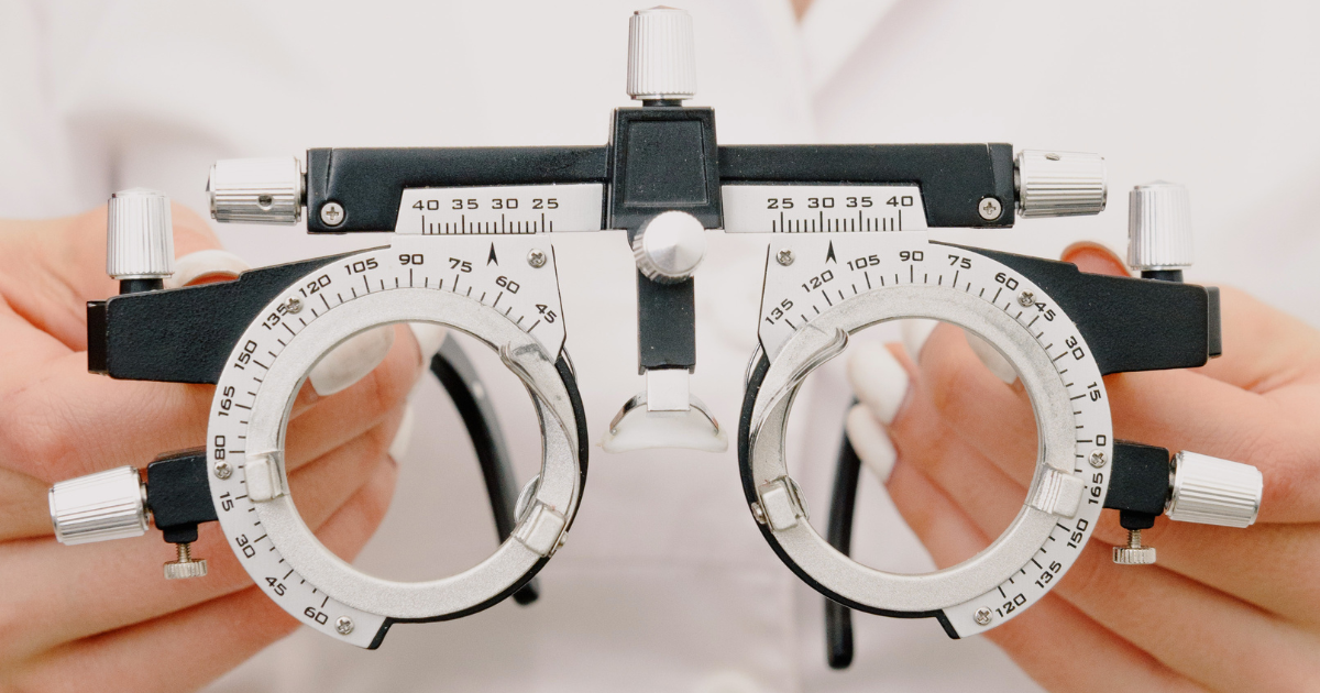
Visual acuity testing measures how clearly each eye sees at a standardized distance. Refraction helps determine whether lenses can improve vision and whether nearsightedness, farsightedness, astigmatism, or presbyopia is present.
The clinician may observe how the pupils respond to light and how the eyes move individually and together. These tests provide information about eye coordination and parts of the neurological visual pathway.
Tonometry measures pressure inside the eye. Pressure is one factor considered when assessing glaucoma risk, but a pressure reading by itself does not confirm or exclude the disease.
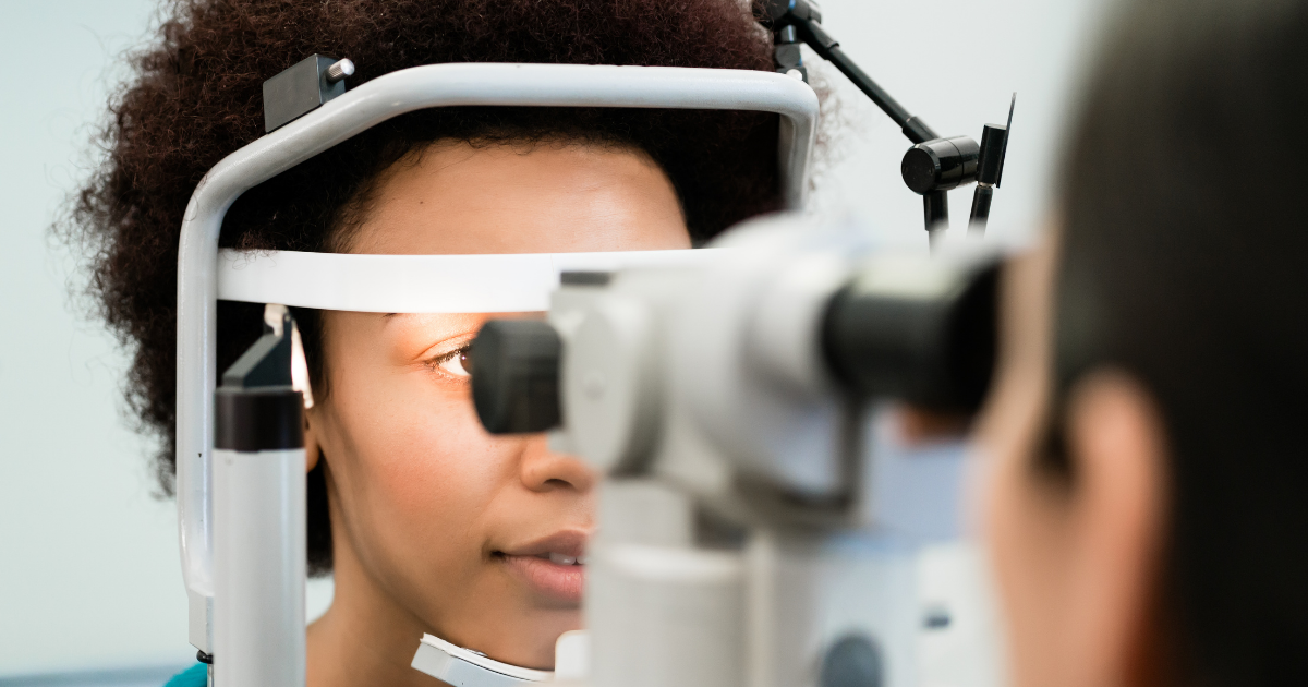
A slit lamp provides a magnified view of structures at the front of the eye, including the eyelids, cornea, iris, and lens. It may help identify inflammation, cataracts, corneal abnormalities, and signs of dry eye.
Examining the back of the eye allows the doctor to evaluate the retina, blood vessels, macula, and optic nerve. The American Academy of Ophthalmology’s eye exam overview explains that specialized tests may also include retinal photography, optical coherence tomography, corneal topography, or visual field testing when clinically appropriate.
Dilating drops temporarily widen the pupil, giving the clinician a broader view of internal eye structures. The examination can help reveal signs of retinal disease, optic nerve damage, diabetic eye disease, and age-related changes.
The National Eye Institute’s guide to dilated examinations describes how dilation supports evaluation for conditions such as glaucoma, cataracts, diabetic retinopathy, and age-related macular degeneration.
After dilation, near vision may remain blurry and the eyes may be more sensitive to light for several hours. Patients should ask whether they will be dilated when scheduling, bring sunglasses, and make transportation arrangements if they do not feel comfortable driving afterward.
Not every appointment requires dilation. The decision depends on risk factors, previous findings, symptoms, and the doctor’s clinical judgment.
Glaucoma is a group of diseases that damage the optic nerve. The most common form often develops gradually, and early peripheral vision loss may not be obvious during daily activities.
The National Eye Institute states that glaucoma symptoms can begin so slowly that a person may not notice them. A comprehensive dilated examination is the way to determine whether the disease is present.
Risk is not determined by eye pressure alone. Clinicians may also consider:
Glaucoma damage cannot simply be reversed with a stronger glasses prescription. Early identification matters because treatment can often slow or prevent additional damage.
Diabetic retinopathy occurs when diabetes damages blood vessels in the retina. Early stages may not cause noticeable symptoms, even though changes are already present.
The Centers for Disease Control and Prevention explains that regular eye examinations can identify diabetes-related problems earlier, when treatment and systemic disease management may help protect sight.
The CDC also recommends that people with diabetes receive yearly comprehensive vision examinations that include dilation. An individual’s medical and eye care teams may recommend a different schedule based on pregnancy, disease severity, previous retinal findings, or other clinical factors.
Managing blood glucose, blood pressure, and cholesterol remains important. Eye treatment addresses ocular complications, but it does not replace broader diabetes care.
The eye provides a direct view of blood vessels and neural tissue. During an examination, a clinician may observe findings associated with conditions such as diabetes, hypertension, inflammatory disease, or medication effects.
This does not mean an eye examination is a universal test for systemic disease. An unusual retinal or optic nerve finding may prompt communication with a primary care clinician or another specialist, who can perform the appropriate medical evaluation.
The CDC’s explanation of why eye exams matter notes that eye doctors may detect signs associated with health problems while examining structures behind the eyes.
There is no ideal schedule for every patient. Recommendations may change based on age, prescription, contact lens wear, medical history, family history, medication use, and previous examination findings.
People may require closer monitoring if they have:
The American Academy of Ophthalmology recommends an eye disease screening by age 40 for adults without known risk factors, while people with symptoms or elevated risk may need evaluation earlier. Existing examination schedules should not be delayed merely because vision seems clear.
A careful history helps the clinician interpret test results. Before an appointment, create a concise list of symptoms, medications, previous eye procedures, and relevant family history.
Describe symptoms precisely. “My vision is blurry” is useful, but “my right eye becomes blurry after 20 minutes of reading and improves after blinking” gives the doctor more diagnostic context.
Routine preventive care is important, but some changes should not wait for the next scheduled visit. Seek prompt professional advice for sudden vision loss, severe eye pain, new double vision, an eye injury, or a sudden increase in flashes and floaters.
A dark curtain or shadow moving across the visual field can be a warning sign of a retinal emergency. New neurological symptoms accompanying vision changes may also require urgent medical evaluation.
When uncertain, contact an eye care or medical professional and describe the symptoms. They can help determine the appropriate level of urgency.
Kittery Optometric Associates provides comprehensive examinations, dry eye care, prescription eyewear, and management of multiple eye health concerns for patients in Kittery and Wells, Maine. The practice also cares for pediatric patients and individuals who need monitoring for conditions such as glaucoma, diabetic eye disease, and retinal disorders.
A normal result offers valuable baseline information for comparison at later visits. An abnormal result creates an opportunity to investigate, monitor, or treat a condition before more vision is affected.
A glasses prescription answers an important question: which lenses produce clearer vision today? A comprehensive examination asks a wider set of questions about how the eyes focus, coordinate, and function, as well as whether their structures appear healthy.
That broader assessment is why symptom-free patients can still benefit from appropriate preventive eye care. Clear vision is reassuring, but it is not conclusive evidence that every part of the visual system is healthy. Examination findings, individual risks, and changes over time provide a much more reliable picture.