Growing Eyes: Children’s Eye Exams in San Juan Capistrano
Children’s eye exams in San Juan Capistrano, CA can evaluate developing visual skills that a brief screening or a child’s ability to read an eye…

Comprehensive eye examinations work by combining several kinds of evidence, not by producing one score that declares the eyes healthy or unhealthy.
This distinction is easy to miss. Patients often remember a prescription number, an eye-pressure reading, or whether they could see the smallest line on a chart. Each result is useful, but each answers a limited question. The clinician’s task is to decide whether the findings support one another, reveal a meaningful change, or require closer investigation.
A comprehensive exam is designed to assess vision, eye function, and ocular structures from different angles. The American Optometric Association’s adult eye examination guideline treats the process as a combination of patient history, visual measurements, ocular function testing, and eye-health evaluation.
Common components may include:
The exact sequence can vary with age, symptoms, medical history, and previous findings. The National Eye Institute’s guide to dilated eye exams similarly describes acuity, visual field, muscle function, pupil response, pressure measurement, and dilation as separate parts of a broader evaluation.
The same result can carry different significance for different patients. A borderline pressure reading means something different in a person with a strong family history of glaucoma than it does in someone with no known risk factors and stable prior findings. A prescription shift may be routine in one setting but prompt additional questions when it appears alongside diabetes, medication changes, or sudden symptoms.
Useful history includes:
Specific descriptions are more useful than saying vision is simply “bad.” Blur that clears after blinking suggests a different line of inquiry from a fixed dark spot or distortion that appears in one eye.
Visual acuity records how clearly a person identifies detail at a particular distance. Refraction tests whether changing lens power improves that clarity. The National Eye Institute explains that myopia, hyperopia, astigmatism, and presbyopia occur when the eye does not focus light correctly on the retina.
If vision improves as expected with refraction, an optical focusing issue may explain the blur. If clarity remains reduced despite an appropriate lens choice, the clinician may investigate other possibilities. Those can involve the cornea, lens, retina, optic nerve, visual pathway, or the reliability of the response itself.
A glasses prescription is therefore one product of an exam, not a summary of the entire exam. Under the Federal Trade Commission’s Eyeglass Rule, prescribers must provide a copy of an eyeglass prescription after a refractive examination without charging an additional fee. That consumer right is separate from the clinical interpretation of eye-health findings.
Clinical measurements are not immune to normal variation. Attention, fatigue, blinking, tear quality, positioning, understanding of the task, and instrument technique can influence a result. A difference from last year may represent a true change, but it may also fall within ordinary measurement variability.
Clinicians look for internal consistency. Does the refraction match the acuity response? Does a visual-field result fit the appearance of the optic nerve? Is an unusual reading repeatable? Do symptoms align with the eye and structure involved?
Repeating a test is not wasted effort. It is one method of checking whether a finding is dependable enough to influence a diagnosis or management plan.
Tonometry provides an important measurement, but there is no single pressure reading that diagnoses or excludes glaucoma for every person. The National Eye Institute’s statement on glaucoma detection explains that pressure testing by itself is insufficient because some people with glaucoma do not have elevated pressure, while others have higher pressure without optic-nerve damage.
A glaucoma assessment may combine pressure with optic-nerve appearance, peripheral vision, corneal characteristics, retinal nerve-fiber measurements, family history, and change over time. The conclusion comes from the pattern, not from treating one number as a universal cutoff.
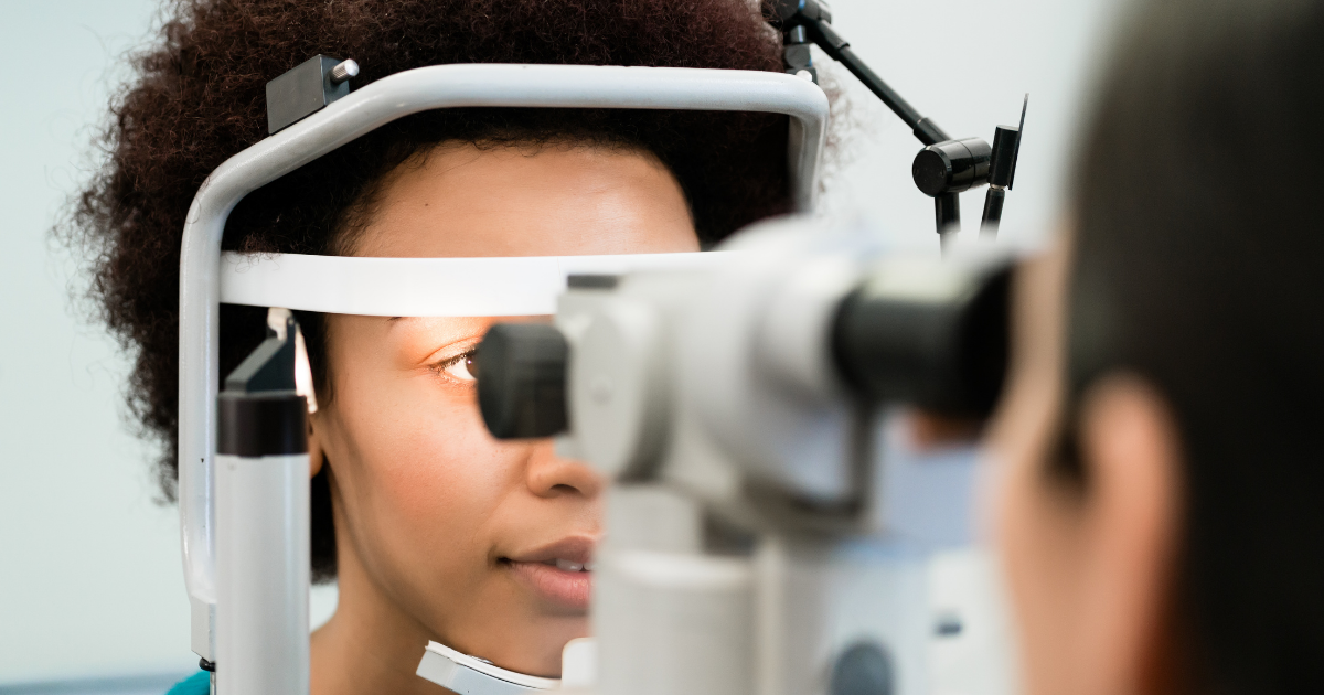
Some tests show how the visual system performs. Others show what eye structures look like. Visual acuity and visual fields are functional measurements. Slit-lamp examination, dilation, ophthalmoscopy, and imaging provide structural information.
The American Academy of Ophthalmology’s eye-exam overview describes the use of multiple techniques to assess vision, pupils, eye movement, pressure, and internal structures. This helps explain why an apparently normal eye-chart result can coexist with an ocular-health concern that has not yet affected central clarity.
Digital retinal images and other scans can document selected structures in detail. They are particularly useful for side-by-side comparison, measurement, and showing patients what the clinician is monitoring.
Images still require context. Image quality can be affected by fixation, pupil size, media clarity, and the area captured. A device may flag a value outside its reference range, but the clinician must decide whether that difference represents disease, anatomy, an artifact, or a result worth repeating.
Technology strengthens an exam when it adds relevant evidence. It should not turn a complex clinical question into a color-coded label without professional interpretation.
A single visit is a snapshot. A series of reliable exams can show whether pressure, prescription, visual fields, retinal appearance, or optic-nerve measurements remain stable. That history helps distinguish a longstanding characteristic from a new development.
Meaningful comparison depends on more than storing data. The clinician must consider whether the same structure was measured, whether test quality was adequate, and whether any change exceeds expected variability. Baseline records become especially valuable when symptoms, health conditions, or treatments change later.
At the end of an exam, findings generally support one or more next steps:
A normal exam does not mean future symptoms should be ignored. Sudden vision loss, new flashes or numerous floaters, a curtain-like shadow, severe pain, significant trauma, or chemical exposure should prompt immediate professional guidance.
Patients can leave with a clearer understanding by asking:
If dilation is expected, bringing sunglasses and planning transportation can make the temporary light sensitivity and near blur easier to manage.
Luminary Eye Care has served Marietta and East Cobb since 2019. Its published comprehensive exam information lists visual acuity, visual fields, retinoscopy, refraction, slit-lamp examination, digital retinal imaging, tonometry, and ophthalmoscopy among the assessments that may be included. The practice states that exams are tailored to each patient’s history and needs.
This combination illustrates why a comprehensive evaluation is more informative than an isolated screening or prescription check. Each test contributes a different piece, while the optometrist determines whether the pieces form a reassuring, uncertain, or concerning pattern.
Georgia consumers can verify optometry licenses through the state’s official professional license lookup.
More testing is not automatically better. The strongest exam is the one that uses appropriate tests, obtains dependable measurements, interprets them in context, and produces a clear next step.
Eye-chart performance, refraction, pressure, fields, structural examination, and imaging each describe one part of vision or eye health. Their real value appears when they are considered together and compared over time. That is how a collection of numbers becomes clinically useful evidence.